NEJM
Review
|
Steven M. Zeitels, M.D., and Gerald B. Healy, M.D.
It has been more than 20 years since Vaughan1 reviewed the state of the art in human laryngology and voice disorders. During that time, an improved understanding of the physiology of voice production has catalyzed medical and surgical interventions, resulting in new procedures, treatments, and opportunities for patients with voice disorders. These advancing initiatives are keeping pace with the aging of the population and the increase in oral communication in the workplace.2
From a person's initial cry to his or her final words, the vocal folds probably sustain the most long-term, soft-tissue trauma of any area of the body. Average conversational pitch frequencies for normal voices are 200 Hz for women and 120 Hz for men, which match the number of collisions per second of the vocal folds and are accompanied by substantial shear stress.
Emotions are manifested primarily in the voice and face, and innervation to both regions is complex. The motor innervation to the vocal muscles is among the densest in the body. It is denser than the facial motor nerve (the seventh cranial nerve), which innervates the muscles of facial expression. The intricacy of laryngeal innervation also allows for one of the most sophisticated human motor skills, high-performance singing.3 The unique nature of singing and oration was revered in many primitive societies. Vocal performance continues to be admired today, as illustrated by the status of religious leaders, educators, entertainers, and, at times, politicians.
A healthy voice is critical for effectiveness at work as well as for psychosocial health. Since most persons, even children, can diagnose the presence of a voice problem during routine conversation, all physicians should be prepared to help patients obtain a proper evaluation. For many people, hoarseness can be disabling and is likely to become more of a problem as voice recognition becomes central to many forms of technology and replaces manual information entry, such as typing and keyboarding.4,5
History
In Europe in 1855, Manuel Garcia,6 an opera teacher, published a report about mirror laryngoscopy. Shortly thereafter, Johann Czermak7 and Ludwig Turck8 modified Garcia's technique for clinical use and helped establish laryngology as a medical specialty. Subsequently, laryngology began to progress as a surgical specialty, owing to improved techniques for treating airway obstruction and vocal disorders. After the Civil War, Jacob Solis-Cohen (Figure 1), who had been a Union general surgeon, became the first specialized head and neck surgeon.9 His extensive achievements include the first long-term cure of a laryngeal cancer10 and the establishment of the first U.S. courses in laryngology.
|

Mirror-guided laryngeal surgery through the oral cavity and pharynx is one of the oldest forms of minimally invasive surgery and was greatly facilitated in the 1880s by the development of mucosal anesthesia with cocaine.11 Indirect laryngoscopy with mirrors was a way to diagnose and treat membranous obstruction of the laryngeal airway. In the 1880s, Joseph O'Dwyer devised tubes for effective cannulation of the larynx.12
Alfred Kirstein formally introduced direct laryngoscopy in 1895.13,14 This technique, combined with cannulation of the larynx,15 led to routine laryngotracheal intubation. Control of the airway by means of orotracheal intubation made possible many subsequent achievements in general anesthesia, cardiopulmonary resuscitation, and critical care medicine.
Phonosurgery
The optimal production of sound from the larynx requires apposition of the edges of the vocal folds (the glottal valve), which are driven into oscillation by the sustained subglottal aerodynamic pressure from the tracheobronchial tree. Ideal vibration requires smooth mucosal edges that close evenly, retaining pliability. Vocal folds function best when they have symmetric rheologic properties and elasticity, which permits the efficient translation of aerodynamic pressure (a power source) into voice (an acoustic signal). The vocal folds are a source of sound, whereas the pharynx, oral cavity, and nose function as a supraglottal resonating chamber, which is a primary component of the human vocal signature.
The vibration of the vocal folds is a fundamental component of voice production. A laryngeal stroboscope synchronizes the frequency of light with the frequency of a person's voice so that the vibration of the vocal folds is visible in slow motion.16,17 Thus, stroboscopy allows the clinician to assess the biomechanical behavior of the microstructure of the vocal folds, which consists of layers of laminae propriae and a layer of epithelium (Figure 2). The widespread use of stroboscopy led to an improved understanding of the pathophysiology of normal and disordered glottal sound production. This understanding of the physiological principles of laryngeal sound, in turn, led to the design of new procedures to correct many voice problems.
|
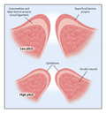
The term "phonosurgery," coined in the early 1960s, refers to surgical procedures that maintain, restore, or enhance the human voice.18,19,20 Phonosurgery includes phonomicrosurgery (endoscopic microsurgery of the vocal folds), laryngoplastic phonosurgery (open-neck surgery that restructures the cartilaginous framework of the larynx and the soft tissues), laryngeal injection (injection of medications as well as synthetic and organic biologic substances), and reinnervation of the larynx.
Phonomicrosurgery
Common Benign Conditions
Glottal lesions may be epithelial in origin (Figure 3), as exemplified by papillomatosis. This viral disease is usually benign but is characterized by foci of invasive carcinoma in extremely rare circumstances. If laryngeal papillomatosis is left untreated, obstruction of the airway will develop in some patients. The disease is most dangerous when it extends caudally, leading to tracheobronchial obstruction and, consequently, urgent palliation of the airway.
|

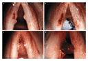
The development of nodules and polyps on the vocal folds (Figure 4) is common because of the repeated collision forces and shear stresses sustained by the delicate subepithelial soft tissues of the vocal folds. These stresses and forces lead to microvascular injury and trauma to the epithelial basement-membrane zone.3,21,22 Overuse or abuse of the vocal cords can lead to vascular malformations such as varices and ectasias,23 which may occur without the presence of a mass lesion. Subepithelial cysts (Figure 5) may be keratinous and congenital in origin but are more frequently mucoid, typically occurring in patients who overuse their voices. In children with lesions, the possibility of a congenital abnormality must always be considered.
|
|
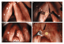
Most nonepithelial lesions result from trauma to the mucosa of the vocal folds that is caused by a patient's abuse or overuse of the voice. This is called "phonotrauma."
A variety of problems involving the laryngeal mucosa are accompanied by laryngopharyngeal reflux of acid and pepsin.24 Reflux causes inflammation of the laryngeal mucosa, which makes the delicate soft tissues more susceptible to phonotrauma as well as to trauma from surgical instruments and intubation. Most patients with laryngopharyngeal reflux do not report having heartburn, which is a symptom of esophagitis. The most common symptoms of laryngopharyngeal reflux are throat clearing and globus sensation (the sensation of having a lump in the throat).
Phonomicrosurgery is used to manage all the aforementioned lesions and is performed through a direct laryngoscope with use of a surgical microscope while the patient is under general anesthesia. The purpose of the surgery is usually to improve the vibratory characteristics of the layered microstructure of the vocal folds. Phonomicrosurgery25,26 has evolved in a stepwise fashion. Shortly after the microscope27 was introduced to laryngology, hand instruments were designed that were suitable for the magnified field. Subsequently, the carbon dioxide laser was introduced28,29,30,31,32 to make hemostatic dissection of soft tissues more precise; it has endured as the most-used type of laser in laryngology.
The drawback of the carbon dioxide laser is that it cannot be used for highly precise tangential cutting without causing heat-induced trauma to the delicate subepithelial tissues of the vocal folds. This photothermal effect leads first to fibrosis and then to varying degrees of hoarseness. Therefore, in the 1990s, there was a return to the use of cold instruments,33 which led to new microflap techniques.25,34,35,36,37 These techniques involve lifting the epithelium of the vocal folds rather than removing it, which preserves the layered microstructure of the vocal folds, the pliability of the mucosa, and, consequently, the production of laryngeal sound. The carbon dioxide laser is still ideal for lesions that are large, vascular, and deeply invasive. Reports of the 585-nm pulsed-dye laser treatment initially introduced by Anderson and Parrish38 for "photoangiolysis"39 of skin lesions suggest that it may be useful for treating a variety of mucosal lesions of the larynx40,41,42 (Figure 4A and Figure 4B) and other types of mucosal disease.
Early Glottic Cancer
Laryngeal cancer, which was rarely noted in the 19th century, has been diagnosed routinely since the introduction of mass-produced cigarettes in the early 20th century.43 By the 1920s precancerous lesions on the vocal folds were becoming common.44 The change from chewing tobacco to smoking coincided with the introduction of antibiotics, which controlled many infectious diseases. As a result, a primary focus of 20th-century laryngology was the optimal management of laryngeal neoplasia.
Most patients in whom early glottic cancer is diagnosed have probably had a lesion on the vocal fold for a number of years, since the cancer typically is preceded by a prolonged period of epithelial dysplasia.44 Despite an adequate resection, dysplasia of the vocal folds frequently recurs, which can be discouraging to both the patient and the surgeon, since the voice usually deteriorates with each intervention. In contrast, phonomicrosurgical resection of early glottic cancer is extremely successful and has evolved from more than a century's worth of endoscopic treatment for laryngeal cancer.45 Cure rates have remained consistent at 90 to 95 percent.46,47,48,49
Hirano et al.50 introduced a graded approach to resection in the 1980s, and since the 1990s ultranarrow resection margins (about 1 to 2 mm)25,26,35,51,52 have been used and have led to improved postoperative voice quality.49 The final vocal outcome has been enhanced further by new techniques of phonosurgical reconstruction, including medialization laryngoplasty53,54 and injection of autogenous fat.55 With the graded approach, nearly normal, conversational-level voices can be achieved, as assessed by objective acoustic and aerodynamic measures.49 Controversy exists regarding the vocal outcome after resection with ultranarrow margins of a T1 glottic cancer, as compared with narrow-field (about 5 cm) irradiation of approximately 66 Gy. With the latter method, it is impossible to avoid administering radiation to the normal tissue of the vocal folds. Radiation-induced stiffness and fibrosis can result, leading to irreparable hoarseness. This can occur in any area of the glottal mucosa, regardless of whether cancer is present.56
Although a multicenter, randomized, controlled study would be necessary to compare the quality of the voice after various treatments for T1a glottic cancer (in which the tumor involves one vocal cord), such a clinical trial is unlikely to occur in the foreseeable future, given the bias of institutions and clinicians and the infrequency of the disease. Regardless of the treatment method, the primary determinant of voice quality is the noncancerous subepithelial superficial lamina propria, since this soft tissue is the principal oscillator. An unanswered question is whether the voice is better after irradiation of the normal and the cancerous tissues of the vocal folds or after a confined microcontrolled resection of portions of the layered microstructure of one diseased vocal fold, with the option of phonosurgical reconstruction.
There is no question that phonomicrosurgery is considerably more cost effective than irradiation,57 since the microlaryngoscopic biopsy includes complete excision, rendering radiation therapy superfluous. With irradiation as the treatment, microlaryngoscopic biopsy and staging are typically the initial components of management. Finally, it is important to consider that irradiation is a valuable single-use treatment for cancer, whereas endoscopic resection can be repeated with minimal morbidity.
Surgical Treatment of Paralysis of the Vocal Folds
Typically, paralysis of the vocal folds results in a breathy and asthenic voice. Patients may have aspiration and dysphagia if the vocal folds are widely abducted or if there is substantial sensory loss in the lower pharynx and larynx. An incompetent or inefficient cough is common and may dramatically exacerbate associated pulmonary and mediastinal disorders.
In the 19th century, electrical stimulation was the sole treatment for paralysis of the vocal folds.58,59,60,61 This approach was based on studies of nerve stimulation in mammals. Throughout most of the 20th century, static reconstruction was accomplished by injection of material into the thyroarytenoid muscle to increase its bulk. In this technique, the medial edge of the paralyzed vocal fold is brought toward the midline to facilitate glottal closure with the neurologically intact musculature of the contralateral cricoarytenoid joint. Such closure permits the aerodynamically induced oscillation of the soft tissues of the vocal folds. Brunings62 described the injection of paraffin for medialization in the early 20th century. In the early 1960s, Teflon63 became the substance of choice because it is available and easy to use.
Today, injection medialization for vocal-fold paralysis is used most frequently to restore glottal competence temporarily. Transitory medialization is often the goal of the treatment, since in many cases paralysis will resolve spontaneously, and which patients will regain normal function is not predictable. Gelfoam64 (gelatin paste) is commonly used for this purpose and is typically absorbed in about three months. Laryngeal electromyography can help clarify whether surgical intervention is necessary, because it helps prognosticate reinnervation.65,66,67
During the past 15 years, repositioning regions of the cartilaginous framework and the underlying soft tissue of the larynx has become the dominant treatment for the dysphonia and dysphagia associated with unilateral vocal-fold paralysis. The repositioned soft tissue of the vocal fold improves the valvular competency of the glottis and therefore sound production. Although this approach was first described at the beginning of the 20th century,68 these procedures were not adopted broadly until they could be performed with the patient under local anesthesia to reestablish aerodynamic glottal competence consistently.69 The operations included implant medialization of the musculomembranous portion of the vocal folds,70,71 which is where vibration occurs for sound emission. For patients who had severe posterior glottal incompetence, a procedure was designed to reestablish the correct adducted arytenoid position posteriorly.72 The need for an arytenoid procedure is based on the severity of posterior incompetence, which is primarily related to reinnervation resulting in idiopathic synkinesis.
A variety of implants73 74 have been described since the initial descriptions of implant medialization. The implants typically vary in density, contour, and adjustability. In the 1990s a new procedure was developed, called adduction arytenopexy, for repositioning the arytenoid.69 The results of adduction arytenopexy closely simulate the normal position of the arytenoid during the production of laryngeal sound. The procedure is performed in combination with the implantation of Gore-Tex lateral to the anterior musculomembranous region75,76 and the alteration of tension by means of cricothyroid subluxation.77 With this approach, normal pitch and dynamic range can be achieved despite complete denervation of one vocal fold.
Laryngoplastic phonosurgical procedures are typically performed while the patient is under local anesthesia with sedation. Local anesthesia is essential, since intraoperative conversation with the patient is necessary to allow for fine adjustments of the framework and soft tissues, which optimize the vocal outcome. Essentially, the paralyzed vocal fold is "tuned" by modifying the height, length, tension, and medial position. Patients have minimal discomfort during the procedure, which is well tolerated even by those who are very ill or have had intracranial or intrathoracic problems. Reestablishing a competent glottal valve can dramatically improve pulmonary toilet for rehabilitation in patients with atelectasis, tracheobronchitis, or pneumonia.
Laryngeal Dystonias
Laryngeal dystonias encompass spasmodic dysphonia and breathing dystonias, the latter being extremely rare.78,79 Spasmodic dysphonia can be understood as inappropriate spasm of the vocal folds during phonation. The spasms may lead to excessive closure (adduction) or opening (abduction). About 85 percent of persons with spasmodic dysphonia have adductor dysfunction, and about 15 percent have abductor dysphonia. Adductor spasmodic dysphonia is often described as a strained and strangled pattern of phonation characterized by inappropriate breaks in sound production. It occurs when excessive closure of the glottal aperture causes vibrations of the vocal folds to cease. Abductor spasmodic dysphonia is a weak and breathy pattern of phonation. It is caused by inadequate closure of the vocal folds, which results in poor vibration secondary to aerodynamic incompetence.
Blitzer et al.79,80 and Ludlow et al.81 championed the use of botulinum toxin A in the treatment of spasmodic dysphonia � a treatment that has revolutionized its management. Small doses of botulinum toxin A are placed in the appropriate intrinsic glottic musculature, typically with electromyographic guidance. Symptoms remit in most patients for a number of months; however, the injections of botulinum toxin must be repeated as symptoms recur.
Berke et al.82 designed a reinnervation procedure in which terminal nerve branches of the adductor musculature are denervated from the recurrent laryngeal nerve and reinnervated by the ansa cervicalis nerve. Although patients often have substantial hypophonia for up to 10 months, Berke and colleagues reported that the procedure is a permanent solution to adductor spasmodic dysphonia. Despite being efficacious, the technique has not gained popularity owing to its complexity.
Pediatric Laryngology
The larynx in a child differs from its adult counterpart in location (it is higher in the neck), consistency (it is softer), and anatomical composition (the connective-tissue layers of the lamina propria of the vocal folds are not well defined). As development progresses, a child's voice matures. Most vocal problems in children result from an abnormality in development.
Dysphonia and hoarseness in children require a thorough assessment, which should include a comprehensive history, obtained from a parent, with specific questions about the neonatal period. A history of respiratory distress at birth, a weak cry, or intubation may all suggest intrinsic laryngeal abnormalities. A careful physical examination is essential. This includes observation of the breathing pattern, as well as auscultation of the larynx for evidence of stridor or an abnormal cry. Videolaryngoscopy with a flexible endoscope can document the appearance and function of the vocal folds and provide a record for further evaluation. Stroboscopy, which is valuable for examining vibration of the vocal folds and thus for evaluating the voice, may be possible in older, cooperative children. However, most young children and all infants require microlaryngoscopy in the operating room under general anesthesia. Furthermore, microlaryngoscopy should be carried out only by experienced pediatric laryngologists and anesthesiologists.
Several disorders are unique to the pediatric larynx. In an infant with dysphonia, a congenital glottic web, vocal-cord paralysis, or congenital cysts may be the causal factor. As children age, nodules on the vocal cords become the most common disorder. These lesions may be caused solely by vocal abuse, but it is possible that gastroesophageal reflux is a cofactor.24 Recurrent respiratory papillomatosis could be the reason for an abnormal voice or abnormal breathing and, if left undiagnosed, may lead to airway obstruction and even death. Therefore, children with hoarse, strained voices should be referred to a pediatric laryngologist for assessment and evaluation. Respiratory papillomatosis may affect a child of any age. Although very rare, even malignant neoplasms can occur in the pediatric larynx. These issues underscore the points that hoarseness in children must not be ignored and that visualization of the vocal folds is imperative when symptoms persist.
Prospects for the Future
Scarred or nonvibratory epithelium in the vocal folds is an important problem. The scarring is often a result of the collision trauma that is associated with abuse of the voice, and it causes mucosal fibrosis and nonpliable epithelium. Scarred vocal-fold membranes are often associated with a variety of lesions, such as polyps, nodules, and cysts. The dysfunction of the vocal folds frequently impairs or terminates the careers of professionals who depend on their voices, such as teachers, executives, politicians, and performing artists.
Scarred epithelium can also result from prolonged endotracheal intubation, the treatment of carcinoma, or laryngotracheal stenosis. A large population of adolescents and adults under the age of 30 years underwent airway reconstruction as infants or children. The elegant procedures, which were developed in the 1970s and modified in the 1980s, allowed these children to function without an artificial airway. However, many of them have some type of vocal dysfunction,83,84 which is typically a result of the placement of life-preserving artificial airways and the subsequent reconstruction of the airways.
It is difficult to determine how many people have dysphonia secondary to nonpliable epithelium, because it is often either unrecognized or considered to be a normal component of the aging voice. However, dysfunctional subepithelial soft tissue is often the result of long-term trauma rather than senescence. Scarred epithelium in the vocal folds can also be a problem for young and middle-aged people with vocally demanding jobs, such as educators, who may subsequently have to leave the workforce. Gray et al.85,86 and Chan et al.87 have been working steadily to characterize the layered microstructure of the vocal folds biochemically and biomechanically. Initiatives in tissue engineering and in the development of biomaterials88 should provide a pliable substitute for the superficial lamina propria in the not-so-distant future.
Another area of fertile research is neurolaryngology. There has been slow but steady progress in selective reinnervation of the intrinsic laryngeal musculature, and a number of research groups in the United States and overseas have devoted substantial resources to this research.89,90,91,92 Trials of the electrical pacing of selected laryngeal muscles in humans have been conducted. It is hoped that selective pacing of the primary abductor of the vocal fold (the posterior cricoarytenoid muscle) will reduce the morbidity associated with bilateral vocal-fold paralysis, which typically leads to airway obstruction and the need for ablative surgery of the vocal folds or tracheotomy. Pacing also provides hope for dynamic treatment of unilateral vocal-fold paralysis, since it could theoretically restore the normal airway aperture; the current state of the art, in contrast, involves the static repositioning of the paralyzed vocal fold and permanent narrowing of the airway.
Strome et al.93 performed a successful larynx transplantation in a person whose larynx was nonfunctional as a result of prior trauma. Most potential candidates for such transplantation, however, require laryngectomy as treatment for cancer and should not undergo transplantation, owing to transplant recipients' concomitant need for immunosuppression. Future methods of minimizing host rejection could make transplantation a viable option, particularly for those people who require only a partial laryngectomy as effective treatment for cancer.
Conclusions
Since its origin, laryngology has been fueled by innovative surgical procedures that improve airway safety as well as the functions of the voice and swallowing. In the past decade there has been a shift in the methods of performing most surgical interventions for vocal dysfunction. Tremendous international collaboration has led to these innovations, which have been achieved with relatively modest funding.
Virtually all physicians converse with their patients and therefore are capable of identifying voice disorders. Most laryngeal lesions exist for years before they are diagnosed, because symptoms typically progress slowly without causing pain or complete disability. Even cancer is preceded by dysplasia for an extended period of time, probably years. Therefore, most disorders remain undiagnosed and untreated at any particular time. Furthermore, hoarseness is often considered to be a normal variant, especially in smokers, or a component of character identification. A person's vocal signature is not always smooth: consider the late Louis Armstrong's voice. Identifying hoarseness is the duty of all physicians and health care professionals. Many medical practitioners care for patients with undiagnosed vocal-fold disorders and discernible hoarseness. A lower threshold for referral for a laryngoscopic evaluation would enhance patient care. Patients should undergo outpatient examination of the larynx if they have unexplained long-term vocal dysfunction or hoarseness of new onset that lasts for more than three weeks.
Laryngeal and voice problems are universal, irrespective of the person's age, sex, or social status. Vocal impairment results in psychosocial problems and loss of productivity in the workplace. The inability to communicate vocally can be crippling to an executive, as well as to a parent, laborer, or performing artist. Care of the larynx requires the cooperation of primary care physicians, laryngeal surgeons, voice scientists, speech pathologists, voice teachers, and family members. Substantial advancements have been made during the past 15 years; this progress should be a harbinger of additional accomplishments in the early 21st century.
Supported by the Advisory Board Foundation, the Eugene B.
Casey Foundation, and the Professor Demetre Souretis Memorial Fund.
Source Information
From the Department of Otology and Laryngology, Harvard Medical School (S.M.Z., G.B.H.), the Division of Laryngology, Massachusetts Eye and Ear Infirmary (S.M.Z.), and the Department of Otolaryngology and Communication Disorders, Children's Hospital (G.B.H.) � all in Boston.
Address reprint requests to Dr. Zeitels at the Division of Laryngology, Massachusetts Eye and Ear Infirmary, 243 Charles St., Boston, MA 02114, or at [email protected].
References
- Vaughan CW. Current concepts in otolaryngology: diagnosis and treatment of organic voice disorders. N Engl J Med 1982;307:863-866. [ISI][Medline]
- Winker MA. Aging in the 21st century: a call for papers. JAMA
2002;287:1326-1326.
[Full Text] - Zeitels SM, Hillman RE, Desloge RB, Mauri M, Doyle PB. Phonomicrosurgery in singers and performing artists: treatment outcomes, management theories, and future directions. Ann Otol Rhinol Laryngol Suppl 2002;111:21-40.
- Kambeyanda D, Singer L, Cronk S. Potential problems associated with use of speech recognition products. Assist Technol 1997;9:95-101. [Medline]
- Haxer MJ, Guinn LW, Hogikyan ND. Use of speech recognition software: a vocal endurance test for the new millennium? J Voice 2001;15:231-236. [ISI][Medline]
- Garcia M. Observations on the human voice. Proc R Soc Lond 1855;7:397-410.
- Czermak JN. Ueber den Kehlkopfspiegel. Wiener Med Wochenschr 1858;8:196-8.
- Turck L. On the laryngeal mirror and its mode of employment, with engravings on wood. Z Gesellschaft Aerzte Wien 1858;26:401-9.
- Zeitels SM. Jacob Da Silva Solis-Cohen: America's first head and neck surgeon. Head Neck 1997;19:342-346. [CrossRef][ISI][Medline]
- Solis Cohen J. Contributions to the clinical history of surgical affections of the larynx. Med Rec 1869;4:244-7.
- Jelinek E. Das Coca�n als An�stheticum und Analgeticum f�r den Pharynx und Larynx. Wien Med Wochenschr 1884;34:1334-7, 1364.
- O'Dwyer J. Chronic stenosis of the larynx treated by a new method, with report of a case. N Y Med Rec 1886;29:641.
- Kirstein A. Autoskopie des Larynx und der Trachea (Laryngoscopia directa, Euthyskopie, Besichtigung ohne Spiegel). Arch Laryngol Rhinol 1895;3:156-64.
- Kirstein A. Autoscopy of the larynx and trachea (direct examination without mirror). Philadelphia: Davis, 1897.
- Elsberg CA. Clinical experiences with intratracheal insufflation (Meltzer), with remarks upon the value of the method for thoracic surgery. Ann Surg 1910;52:23-29.
- Oertel M. Das Laryngo-Stroboskop und die laryngo-stroboskpische Untersuchung. Arch Laryngol Rhinol 1895;3:1-16.
- von Leden H. The electronic synchron-stroboscope: its value for the practicing laryngologist. Ann Otol Rhinol Laryngol 1961;70:881-893. [ISI]
- von Leden H. Plastic surgery of the larynx. Rev Panam Otorrinolaringol Broncoesofagol 1963;1:7-11.
- von Leden H. Surgery for the improvement of vocal function. Rev Panam Otorrinolaringol Broncoesofagol 1969;3:137-43.
- von Leden H. The history of phonosurgery. In: Ford CN, Bless DM, eds. Phonosurgery: assessment and surgical management of voice disorders. New York: Raven Press, 1991:3-24.
- Gray S. Basement membrane zone injury in vocal nodules. Vocal fold physiology conference: acoustic, perceptual, and physiological aspects of voice mechanisms. San Diego: Singular, 1991:21-7.
- Gray SD, Hammond E, Hanson DF. Benign pathologic responses of the larynx. Ann Otol Rhinol Laryngol 1995;104:13-18. [ISI][Medline]
- Hochman II, Sataloff RT, Hillman RE, Zeitels SM. Ectasias and varices of the vocal fold: clearing the striking zone. Ann Otol Rhinol Laryngol 1999;108:10-16. [ISI][Medline]
- Koufman JA. The otolaryngologic manifestations of gastroesophageal reflux disease (GERD). Laryngoscope 1991;101:Suppl 53:1-78. [ISI][Medline]
- Zeitels SM. Premalignant epithelium and microinvasive cancer of the vocal fold: the evolution of phonomicrosurgical management. Laryngoscope 1995;105:1-51.
- Zeitels SM. Atlas of phonomicrosurgery and other endolaryngeal procedures for benign and malignant disease. San Diego: Singular, 2001.
- Scalco AN, Shipman WF, Tabb HG. Microscopic suspension laryngoscopy. Ann Otol Rhinol Laryngol 1960;69:1134-1138.
- Jako GJ. Laser surgery of the vocal cords: an experimental study with carbon dioxide lasers on dogs. Laryngoscope 1972;82:2204-2216. [ISI][Medline]
- Strong MS, Jako GJ. Laser surgery of the larynx: early clinical experience with continuous CO2 laser. Ann Otol Rhinol Laryngol 1972;81:791-798. [ISI][Medline]
- Strong MS. Laser excision of carcinoma of the larynx. Laryngoscope 1975;85:1286-1289. [ISI][Medline]
- Vaughan CW. Transoral laryngeal surgery using the CO2 laser: laboratory experiments and clinical experience. Laryngoscope 1978;88:1399-1420. [ISI][Medline]
- Vaughan CW, Strong MS, Jako GJ. Laryngeal carcinoma: transoral treatment using the CO2 laser. Am J Surg 1978;136:490-493. [ISI][Medline]
- Zeitels SM. Laser versus cold instruments for microlaryngoscopic surgery. Laryngoscope 1996;106:545-552. [ISI][Medline]
- Bouchayer M, Cornut G. Instrumental microscopy of benign lesions of the vocal folds. In: Ford CN, Bless DM, eds. Phonosurgery: assessment and surgical management of voice disorders. New York: Raven, 1991:143-65.
- Zeitels SM. Microflap excisional biopsy for atypia and microinvasive glottic cancer. Operative Techniques Otolaryngol Head Neck Surg Phonosurg Ed 1993;4:218-22.
- Sataloff R, Spiegel JR, Heuer RJ, et al. Laryngeal mini-microflap: a new technique and reassessment of the microflap saga. J Voice 1995;9:198-204. [ISI][Medline]
- Courey MS, Gardner GM, Stone RE, Ossoff RH. Endoscopic vocal fold microflap: a three-year experience. Ann Otol Rhinol Laryngol 1995;104:267-273. [ISI][Medline]
- Anderson RR, Parrish JA. Selective photothermolysis: precise microsurgery by selective absorption of pulsed radiation. Science 1983;220:524-527. [ISI][Medline]
- Anderson RR, Jaenicke KF, Parrish JA. Mechanisms of selective vascular changes caused by dye lasers. Lasers Surg Med 1983;3:211-215. [Medline]
- Bower CM, Waner M, Flock S, Schaeffer R. Flash pump dye laser treatment of laryngeal papillomas. Ann Otol Rhinol Laryngol 1998;107:1001-1005. [ISI][Medline]
- McMillan K, Shapshay SM, McGilligan JA, Wang Z, Rebeiz EE. A 585-nanometer pulsed dye laser treatment of laryngeal papillomas: preliminary report. Laryngoscope 1998;108:968-972. [ISI][Medline]
- Franco RA Jr, Zeitels SM, Farinelli WA, Anderson RR. 585-nm Pulsed dye laser treatment of glottal papillomatosis. Ann Otol Rhinol Laryngol 2002;111:486-492. [ISI][Medline]
- Castro J. Tobacco Road's dirty ashtrays: an heir to a cigarette fortune portrays a grim family legacy. Time. April 24, 1989:52.
- Jackson C. Cancer of the larynx: is it preceded by a recognizable precancerous condition? Trans Am Laryngol Assoc 1922;44:182-201.
- Fr�nkel B. Erste Helung eines Larynx-Cancoids vermittelst Ausrottung per vias naturales. Arch Klin Chir 1886;34:281-6.
- DeSanto LW. Selection of treatment for in situ and early invasive carcinoma of the glottis. In: Alberti PW, Bryce DB, eds. Workshops from the Centennial Conference on Laryngeal Cancer. New York: Appleton-Century-Crofts, 1976:146-50.
- Blakeslee D, Vaughan CW, Shapshay SM, Simpson GT, Strong MS. Excisional biopsy in the selective management of T1 glottic cancer: a three-year follow-up study. Laryngoscope 1984;94:488-494. [ISI][Medline]
- Steiner W. Results of curative laser microsurgery of laryngeal carcinomas. Am J Otolaryngol 1993;14:116-121. [ISI][Medline]
- Zeitels SM, Hillman RE, Franco RA, Bunting GW. Voice and treatment outcome from phonosurgical management of early glottic cancer. Ann Otol Rhinol Laryngol Suppl 2002;111:1-20.
- Hirano M, Hirade Y, Kawasaki H. Vocal function following carbon dioxide laser surgery for glottic carcinoma. Ann Otol Rhinol Laryngol 1985;94:232-235. [ISI][Medline]
- Zeitels SM. Phonomicrosurgical treatment of early glottic cancer and carcinoma in situ. Am J Surg 1996;172:704-709. [CrossRef][ISI][Medline]
- Remacle M, Lawson G, Jamart J, Minet M, Watelet JB, Delos M. CO2 laser in the diagnosis and treatment of early cancer of the vocal fold. Eur Arch Otorhinolaryngol 1997;254:169-176. [ISI][Medline]
- Zeitels SM, Jarboe J, Franco RA. Phonosurgical reconstruction of early glottic cancer. Laryngoscope 2001;111:1862-1865. [ISI][Medline]
- Remacle M, Lawson G, Hedayat A, Trussart C, Jamart J. Medialization framework surgery for voice improvement after endoscopic cordectomy. Eur Arch Otorhinolaryngol 2001;258:267-271. [ISI][Medline]
- Zeitels SM. Glottic reconstruction and voice rehabilitation. Atlas of phonomicrosurgery and other endolaryngeal procedures for benign and malignant disease. San Diego: Singular, 2001:219-21.
- Lehman JJ, Bless DM, Brandenburg JH. An objective assessment of voice production after radiation therapy for stage I squamous cell carcionoma of the glottis. Otolaryngol Head Neck Surg 1988;98:121-129. [ISI][Medline]
- Myers EN, Wagner RL, Johnson JT. Microlaryngoscopic surgery for T1 glottic lesions: a cost-effective option. Ann Otol Rhinol Laryngol 1994;103:28-30. [ISI][Medline]
- Hooper FH. Experimental researches on the tension of the vocal bands. Trans Am Laryngol Assoc 1883;5:118-137.
- Hooper FH. Concerning the positions of the paralyzed vocal bands. Trans Am Laryngol Assoc 1886;8:22-33.
- Motor paralysis of the larynx. In: Solis-Cohen J. Diseases of the throat: a guide to the diagnosis and treatment. 2nd ed. New York: William Wood, 1879:636-59.
- Laryngeal paralysis from disease or injury of the medulla oblongata. In: Mackenzie M. Diseases of the pharynx, larynx, and trachea. New York: William Wood, 1880:309-28.
- Technique of autoscopic operations. In: Brunings W. Direct laryngoscopy, bronchoscopy, and esophagoscopy. London: Bailliere, Tindall, & Cox, 1912:116-20.
- Arnold GE. Vocal rehabilitation of paralytic dysphonia. VI. Further studies of intracordal injection materials. Arch Otolaryngol 1961;73:290-294.
- Schramm VL, May M, Lavorato AS. Gelfoam paste injection for vocal cord paralysis: temporary rehabilitation of glottic incompetence. Laryngoscope 1978;88:1268-1273. [ISI][Medline]
- Ford C. Laryngeal EMG in clinical neurolaryngology. Arch
Otolaryngol Head Neck Surg 1998;124:472-475.
[Full Text] - Woodson GE. Clinical value of laryngeal EMG is dependent on
experience of the clinician. Arch Otolaryngol Head Neck Surg
1998;124:476-476.
[Full Text] - Koufman JA, Postma GN, Whang CS, et al. Diagnostic laryngeal electromyography: the Wake Forest experience 1995-1999. Otolaryngol Head Neck Surg 2001;124:603-606. [CrossRef][ISI][Medline]
- Payr A. Plastik am Schildknorpel zur Behebung der Folgen einseitiger Stimmbandlahmung. Dtsch Med Wochenschr 1915;43:1265-1270.
- Zeitels SM, Hochman I, Hillman RE. Adduction arytenopexy: a new procedure for paralytic dysphonia with implications for implant medialization. Ann Otol Rhinol Laryngol 1998;107:2-24.
- Isshiki N, Morita H, Okamura H, Hiramoto M. Thyroplasty as a new phonosurgical technique. Acta Otolaryngol (Stockh) 1974;78:451-457. [ISI][Medline]
- Koufman JA. Laryngoplasty for vocal cord medialization: an alternative to Teflon. Laryngoscope 1986;96:726-731. [ISI][Medline]
- Isshiki N, Tanabe M, Sawada M. Arytenoid adduction for unilateral vocal cord paralysis. Arch Otolaryngol 1978;104:555-558. [Abstract]
- Montgomery WW, Blaugrund SM, Varvares MA. Thyroplasty: a new approach. Ann Otol Rhinol Laryngol 1993;102:571-579. [ISI][Medline]
- Netterville JL, Stone RE, Civantos FJ, Ossoff RH. Silastic medialization and arytenoid adduction: the Vanderbilt experience. Ann Otol Rhinol Laryngol 1993;102:413-424. [ISI][Medline]
- McCulloch TM, Hoffman HT. Medialization laryngoplasty with expanded polytetrafluoroethylene: surgical technique and preliminary results. Ann Otol Rhinol Laryngol 1998;107:427-432. [ISI][Medline]
- Zeitels SM, Mauri M, Dailey SH. Medialization laryngoplasty with Gore-Tex for voice restoration secondary to glottal incompetence: indications and observations. Ann Otol Rhinol Laryngol 2003;112:180-184. [ISI][Medline]
- Zeitels SM, Hillman RE, Desloge RB, Bunting GA. Cricothyroid subluxation: a new innovation for enhancing the voice with laryngoplastic phonosurgery. Ann Otol Rhinol Laryngol 1999;108:1126-1131. [ISI][Medline]
- Grillone GA, Blitzer A, Brin MF, Annino DJ Jr, Saint-Hilaire MH. Treatment of adductor laryngeal breathing dystonia with botulinum toxin type A. Laryngoscope 1994;104:30-32. [ISI][Medline]
- Blitzer A, Brin MF, Stewart CF. Botulinum toxin management of spasmodic dysphonia (laryngeal dystonia): a 12-year experience in more than 900 patients. Laryngoscope 1998;108:1435-1441. [ISI][Medline]
- Blitzer A, Brin MF, Fahn S, Lovelace RE. Clinical and laboratory characteristics of focal laryngeal dystonia: study of 110 cases. Laryngoscope 1988;98:636-640. [ISI][Medline]
- Ludlow CL, Naunton RF, Sedory SE, Schulz GM, Hallett M. Effects of botulinum toxin injections on speech in adductor spasmodic dysphonia. Neurology 1988;38:1220-1225. [Abstract]
- Berke G, Blackwell KE, Gerratt BR, Verneil A, Jackson KS, Sercarz JA. Selective laryngeal adductor denervation-reinnervation: a new surgical treatment for adductor spasmodic dysphonia. Ann Otol Rhinol Laryngol 1999;108:227-231. [ISI][Medline]
- MacArthur CJ, Kearns GH, Healy GB. Voice quality after laryngotracheal reconstruction. Arch Otolaryngol Head Neck Surg 1994;120:641-647. [Abstract]
- Smith ME, Marsh JH, Cotton RT, Myer CM III. Voice problems after pediatric laryngotracheal reconstruction: videolaryngostro-boscopic, acoustic, and perceptual assessment. Int J Pediatr Otorhinolaryngol 1993;25:173-181. [ISI][Medline]
- Gray SD, Titze IR, Chan R, Hammond TH. Vocal fold proteoglycans and their influence on biomechanics. Laryngoscope 1999;109:845-854. [ISI][Medline]
- Gray SD, Titze IR, Alipour F, Hammond TH. Biomechanical and histological observations of vocal fold fibrous proteins. Ann Otol Rhinol Laryngol 2000;109:77-85. [Medline]
- Chan RW, Gray SD, Titze IR. The importance of hyaluronic acid in vocal fold biomechanics. Otolaryngol Head Neck Surg 2001;124:607-614. [CrossRef][ISI][Medline]
- Niklason LE, Langer R. Prospects for organ and tissue
replacement. JAMA 2001;285:573-576.
[Abstract/Full Text] - Broniatowski M. Bionic larynx: electronic control of the reimplanted organ in the dog. Laryngoscope 1988;98:1107-1115. [ISI][Medline]
- Kojima H, Omori K, Nonomura M, Honjo I, Isshiki N, Shimizu Y. Electrical pacing for dynamic treatment of unilateral vocal cord paralysis: experiment in long-denervated muscle. Ann Otol Rhinol Laryngol 1991;100:15-18. [ISI][Medline]
- Goldfarb D, Keane WM, Lowry LD. Laryngeal pacing as a treatment for vocal fold paralysis. J Voice 1994;18:179-185.
- Zealear DL, Rainey CL, Herzon GD, Netterville JL, Ossoff RH. Electrical pacing of the paralyzed human larynx. Ann Otol Rhinol Laryngol 1996;105:689-693. [ISI][Medline]
- Strome M, Stein J, Esclamado R, et al. Laryngeal
transplantation and 40-month follow-up. N Engl J Med 2001;344:1676-1679.
[Full Text]