UK
Europe
USA&Canada
Aust.&NZ
Asia
UK
Europe
USA&Canada
Aust.&NZ
Asia
Editorials Online�
Medical
Law News
E-texts
Links
Classifieds
Home
|
|
|
NEJM
Jerry R. Mendell, M.D., and Zarife Sahenk, M.D., Ph.D.
This Journal feature begins with a case vignette highlighting a common clinical problem. Evidence supporting various strategies is then presented, followed by a review of formal guidelines, when they exist. The article ends with the authors' clinical recommendations. A 67-year-old woman who had been in excellent health noticed the onset of burning pain in the left great toe two years before evaluation. The pain subsequently extended to involve both feet, from the toes to the heels, and was associated with numbness, tingling, and burning. The discomfort has become severe, is present throughout the day, and disrupts sleep. A physical examination reveals normal muscle strength, muscle-stretch reflexes, proprioception, and vibratory sensation; only pinprick sensation in the toes and feet is diminished. How should this patient be evaluated and treated? The Clinical Problem There are many causes of painful sensory neuropathy (Table 1). In one subtype referred to as "small-fiber painful sensory neuropathy," only the A- (small myelinated) and nociceptive C (unmyelinated) nerve fibers are affected. Studies indicate that this condition represents the most common type of painful sensory neuropathy in patients older than 50 years of age. It is vastly underrecognized, and in most cases, no cause can be found.1,2,3 In another group of neuropathies associated with pain, the discomfort is caused in part by damage to small nerve fibers, but large nerve fibers (A- and A- nerve fibers) that are responsible for proprioception, vibratory sensation, muscle-stretch reflexes, and muscle strength are also affected. The distinction between the two subtypes of painful sensory neuropathies is not trivial, since the underlying cause is more likely to be identifiable when both large and small fibers are affected.1 Irrespective of the subtype of neuropathy, the pain generated by damage to small nerve fibers is debilitating and responds poorly to treatment. Finding and treating the cause is the best long-term strategy but is not routinely possible, and even when it is possible, treatment may not begin to relieve pain for many months or longer.
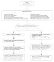
Strategies and Evidence Initial Evaluation Since neuropathy is not the only cause of pain in the feet, one must first determine whether the peripheral nerve is the source of discomfort. Typical symptoms of neuropathic pain related to small fibers include burning (the sensation that the feet are on fire), sharp pain (described as knife-like, jabbing, or pins and needles), shooting pain, and aching in the toes and feet (reflecting damage to the longest axons). Pain emanating from the peripheral nerves is indicated by the description of the feet as tingling, numb, or feeling tight, wooden, or dead. Peripheral-nerve pain is often exacerbated at night, but some patients describe pressure-induced pain with standing or walking. The history will help distinguish among problems associated with plantar fasciitis, arthritis, bursitis, tendonitis, and polymyalgia rheumatica.4 Lumbosacral radiculopathies (with or without spinal stenosis) are not dependent on nerve length and may be accompanied by paraspinal muscle spasm and aggravated by activities (such as lifting). Pain in the toes, related to entrapment of the posterior tibial nerve at the tarsal tunnel (the space beneath the flexor retinaculum and behind the medial malleolus), may mimic painful sensory neuropathy.5 Nerve entrapment at the carpal tunnel accompanying painful sensory neuropathy may point to diabetes mellitus or amyloidosis. In disorders with exclusive or predominant involvement of small nerve fibers, there is a dramatic mismatch between symptoms and observable neurologic deficits. In the typical small-fiber sensory neuropathy affecting patients older than 50 years of age,1 there is an abnormal loss of pinprick sensation in the feet, which may extend centripetally to the level of the knees but rarely above the knees. The sensation of touch may also be diminished, whereas other types of sensation are preserved. In painful sensory neuropathies affecting both large and small fibers, there is reduced proprioception, loss of muscle-stretch reflexes, and muscle weakness, reflecting the loss of large fibers. Loss of vibratory sensation that is restricted to the toes can be a normal finding in the elderly but is abnormal if it extends to the ankles. Two findings on physical examination may help distinguish the pain of tarsal tunnel syndrome from small-fiber neuropathy: Tinel's sign (tingling in the limb served by the nerve after percussion) over the tarsal tunnel and tenderness to palpation over the flexor retinaculum.5 A loss of sensation that is restricted to the medial aspect of the foot, sparing the heel, also points to tarsal tunnel syndrome. The initial evaluation must include electromyography and nerve-conduction studies, unless the diagnosis is known (for example, in a patient with diabetes and known microvascular disease). Electrodiagnostic studies are useful in patients with painful sensory neuropathy for identifying a mononeuropathy (such as focal entrapment at the tarsal tunnel); differentiating multiple mononeuropathy (which is characteristic of peripheral-nerve vasculitis) from polyneuropathy (which is symmetric); and distinguishing axonal neuropathies (e.g., diabetic neuropathy) from demyelinating neuropathies.6 Normal studies are consistent with pure small-fiber neuropathy. Laboratory evaluation should be guided by the results of electrodiagnostic testing (Figure 1). If electrodiagnostic studies are normal, nonneuropathic causes of pain (including local inflammation, such as arthritis or plantar fasciitis, or central nervous system causes, such as myelopathy) must be considered; further testing is warranted to establish the diagnosis of small-fiber neuropathy. The sudomotor-axon reflex test, which quantitates sweating, is a practical, highly specific, and sensitive method (sensitivity, approximately 80 percent) for documenting damage to small nerve fibers.7 Skin biopsies that demonstrate loss of intraepidermal nerve fibers represent an alternative method with slightly greater sensitivity for documenting small-fiber neuropathy: approximately 10 percent of patients with a normal sweat test will have abnormal skin biopsies.1,2 However, skin biopsies are not widely available, and the morphometric analysis is laborious. Quantitative sensory testing assesses small-fiber damage by measuring pain and temperature thresholds in the skin.3 Sensitivity and specificity are lower than those of skin biopsies or sudomotor testing,1,3 and performance depends on patients' cooperation and attention.8
Treatment of Painful Neuropathies Management of the neuropathy is guided by two principles: treatment of the underlying condition (which will not be discussed here) and strategies designed to relieve peripheral-nerve pain irrespective of cause. Pathophysiology of Painful Neuropathy Pain is a protective response to tissue injury, but persistent pain is maladaptive. Pain can occur without provocation (be stimulus-independent, as with burning and paresthesias accompanying small-fiber neuropathies) or can be stimulus-evoked (for example, hyperalgesia in response to noxious stimuli or allodynia induced by non-noxious stimuli). The cause of the nerve damage does not dictate the type of pain, and nonspecific therapies that are effective for one cause should also be applicable to others. Figure 2 summarizes the pathophysiology of pain from peripheral neuropathy and suggests potential pharmacologic strategies for treatment.
Summary of Clinical Trials Judging the efficacy of treatments for painful neuropathies is challenging. Reports can be misleading, because results for a given drug can be statistically significant despite the fact that good or excellent pain relief has been achieved in relatively few patients. In addition, patients expect substantial pain relief with relatively few side effects. Failure to meet these expectations leads to disappointment. We summarize here the results of randomized, controlled trials of agents for painful sensory neuropathy. Although the "number needed to treat" (an estimate of the total number of patients who would need to be treated in order to achieve 50 percent pain relief in one patient) has merits, because it provides information on both the rate and the magnitude of response,9 it also has limitations, especially when it is used to compare studies performed in different populations of patients or for different durations. Thus, we provide the size of the treated cohort and the percentage of cohort members who have a response to treatment. Antidepressant Drugs Tricyclic Antidepressants No agents have been as thoroughly studied for relief of neuropathic pain as the tricyclic antidepressants.10 These drugs block reuptake of serotonin and noradrenaline and presumably relieve pain by inhibition of the sodium channel. Both spontaneous pain and hyperalgesia respond to tricyclic agents. Approximately 300 subjects with diabetic neuropathy have participated in controlled trials of various tricyclic agents. The accumulative efficacy suggests that about one third of patients achieve a 50 percent reduction in neuropathic pain.11,12,13 Responses are often insufficient in clinical practice, and benefits are sometimes outweighed by side effects, especially among the elderly (Table 2).
Selective Serotonin-Reuptake Inhibitors Selective serotonin-reuptake inhibitors differ from tricyclic antidepressants in that they selectively block serotonin reuptake. Clinical trials of these agents (which have involved fewer than 100 patients overall) suggest that their efficacy is lower than that of tricyclic agents.14,15,16 Paroxetine reduces the pain of diabetic neuropathy better than placebo but was not as effective as the tricyclic antidepressant imipramine in a head-to-head comparison.14 Citalopram diminishes neuropathic pain with an efficacy equal to that of paroxetine,15 whereas fluoxetine showed no benefit in diabetic neuropathy.16 Other Antidepressants Venlafaxine has fewer side effects than typical tricyclic antidepressants because of reduced binding to muscarinic, histamine, and 1-adrenergic receptors; one small randomized study suggested that the drug had benefit in patients with painful sensory neuropathy related to cancer.17 Bupropion, a second-generation, specific inhibitor of neuronal norepinephrine reuptake, diminished neuropathic pain by about 30 percent in a cohort of 41 subjects with neuropathy from multiple causes who were treated for six weeks.18 Anticonvulsants Carbamazepine Carbamazepine stabilizes membranes by inhibiting sodium channels. Although it is effective for trigeminal neuralgia, data with regard to painful peripheral neuropathy are limited. One placebo-controlled trial involving 30 subjects19 suggested a benefit in diabetic neuropathy equivalent to that of tricyclic antidepressants. In practice, intolerance to the side effects of carbamazepine limits its use, especially in the elderly. Oxcarbazepine, a keto-acid analogue of carbamazepine, is better tolerated. Data on the drug's efficacy for painful sensory neuropathy are not available, but its efficacy for trigeminal neuralgia is similar to that of carbamazepine.20 Phenytoin Phenytoin, which also blocks sodium channels, is rarely used as first-line therapy for neuropathic pain, since it has inconsistent effectiveness in patients with painful diabetic neuropathy.21,22 However, possible benefit was suggested by a recent small study reporting a reduction in pain due to neuropathy from various causes after a single intravenous infusion of phenytoin.23 Gabapentin Gabapentin was designed as a -aminobutyric�acid agonist, but its precise mechanism of action remains uncertain. Two clinical trials demonstrated pain relief in patients with diabetic neuropathy,24,25 whereas a third trial did not.26 When compared head-to-head with amitriptyline, gabapentin had equal efficacy.25 Reduction in neuropathic pain required doses higher than 1600 mg per day � an important consideration, since many patients are given doses that are too small. The side-effect profile of gabapentin is more favorable than those of many other agents, but nearly 25 percent of patients report dizziness, and 30 percent report sedation. Lamotrigine Lamotrigine (at a dose of 400 to 600 mg per day) resulted in moderate pain relief with minimal side effects in a single small trial involving patients with diabetic or human immunodeficiency virus (HIV)�associated neuropathy.27 Antiarrhythmic Drugs Mexiletine Intravenous lidocaine produced moderate reductions in pain in patients with diabetic neuropathy, but this method of administration is impractical.28 There have been inconsistent results with the use of mexiletine, the oral analogue of lidocaine. Two studies in patients with diabetic neuropathy showed a beneficial effect29,30; another demonstrated efficacy with regard to secondary outcomes but not with regard to global pain relief31 ; and a fourth trial in patients with diabetic neuropathy,32 as well as a trial in patients with HIV-associated neuropathy,33,34 failed to demonstrate benefit. N-Methyl-D-Aspartate Glutamate Antagonists Very few controlled studies, involving only a small number of patients with diabetic neuropathy, have addressed the efficacy of N-methyl-D-aspartate glutamate antagonists (e.g., dextromethorphan) in painful sensory neuropathy.35,36 The studies suggest a beneficial effect in selected patients who can tolerate the sedation, but there are numerous side effects, including impairment of memory, ataxia, and motor incoordination. Narcotic and Nonnarcotic Analgesics Clinicians whose patients have refractory painful sensory neuropathy may feel pressure to use opioid analgesics, although there is concern about the potential for addiction. Oxycodone has been shown to reduce pain in postherpetic neuralgia,37 but data are sparse regarding the effects of opioid analgesics on painful sensory neuropathy. An article in this issue of the Journal38 demonstrates that the opioid agonist levorphanol reduced neuropathic pain (including pain in 32 patients with sensory neuropathy) by 36 percent, at an average daily dose of 8.9 mg. However, side effects were frequent � including itching, mood changes, weakness, and confusion. These side effects were less common when lower doses were used, but lower doses were less effective. Efficacy was lower for painful sensory neuropathy than for postherpetic neuralgia, spinal cord injury, or multiple sclerosis, underscoring the refractory nature of pain from damage to peripheral nerves. Tramadol is a drug that shares properties with opioid analgesics but demonstrates low-affinity binding to �-opioid receptors. It is well tolerated and less likely than other opioid agonists to cause dependence and lead to abuse. Data from trials involving approximately 100 patients with painful sensory neuropathy related to diabetes or other causes39,40 suggest that the efficacy of tramadol is similar to that of tricyclic antidepressants or levorphanol. Nausea and constipation occur in about 20 percent of patients, and headache and somnolence occur in about 15 percent, but generally the drug is well tolerated. Levodopa Dopamine agonists can modify pain, presumably through the inhibition of input to segments of the spinal cord. A single study demonstrated a reduction of pain in a small cohort of subjects with diabetic neuropathy.41 Topical Agents Capsaicin Capsaicin depletes substance P from sensory nerves in the skin, but outcomes in patients with neuropathy have been inconsistent. At least three studies involving more than 250 subjects in total have shown moderate efficacy in diabetic neuropathy.42,43,44 In contrast, no pain relief was achieved in patients with chronic painful distal neuropathy or HIV-associated neuropathy.45,46 In practice, the effects of capsaicin are inconsistent, and a disincentive to use it is that pain is exacerbated when it is first administered. Topical Lidocaine Topically applied lidocaine exerts effects by reducing ectopic neural discharges in superficial nerves. Patches containing 5 percent lidocaine have been approved by the Food and Drug Administration for postherpetic neuralgia. In peripheral neuropathies, the pain extends over wider areas, which limits the usefulness of such patches, but some patients may benefit from patches trimmed to match a specific area where there is excessive pain. Alternative Therapies In the only controlled study of acupuncture for peripheral-nerve pain related to HIV, the placement of needles in traditional sites resulted in no greater relief of pain than the use of sham sites.47 Although transcutaneous stimulation of nerves showed short-term benefit among subjects with diabetic neuropathy,48,49 it has not been effective in practice. Areas of Uncertainty At best, current therapies for painful sensory neuropathy result in a 30 to 50 percent reduction in pain, and such a reduction rarely meets patients' expectations. Randomized trials are warranted for established anticonvulsant agents (such as valproic acid and clonazepam) as well as the newer anticonvulsant agents49 (such as oxcarbazepine, tiagabine,50 topiramate, pregabalin, and vigabatrin). The antidepressants venlafaxine and bupropion also merit additional study. It remains uncertain whether adequate pain relief can be achieved with a multidrug strategy, particularly with the use of pharmacologic agents targeted at more than one site in the pain pathway. Guidelines There are no guidelines available from professional organizations for the treatment of painful sensory neuropathy. Summary and Conclusions Treatment of painful sensory neuropathy presents enormous challenges and is currently inadequate. The evaluation of patients with this condition does not necessarily require a neurologist, but it does require clinicians experienced with electromyography and autonomic nervous system testing (Figure 1). Education of the patient is critical in order to define realistic goals and expectations. Patients must understand that complete relief of pain is unlikely to be achieved with our current armamentarium of agents (Table 2). A diary of side effects and perceived benefits should be maintained by patients and shared with the physician so that drug regimens can be adjusted as necessary. Because monotherapy generally results in a 30 to 50 percent reduction in pain at best, a multidrug regimen may be helpful. Although data are lacking to support the use of combination therapy, a logical strategy is to use combinations of drugs that target different sites in the pain pathway (Figure 2). There is no one set approach to the treatment of patients with painful sensory neuropathy such as the patient described in the vignette. We consider gabapentin to be a reasonable first choice on the basis of clinical trials showing efficacy and its relatively favorable side-effect profile. A starting dose of 900 mg per day is well tolerated, but in all probability, higher doses will be necessary. The dose should be slowly increased to at least 1600 mg per day and can be as high as 3600 mg per day, if necessary. If pain relief is inadequate at the maximal dose, then another drug should be added and its dose slowly increased. Tramadol has shown efficacy in clinical trials and is also well tolerated; we would therefore add this agent in patients who have inadequate pain relief with gabapentin alone and would substitute tramadol for gabapentin in patients who are intolerant of gabapentin. If pain persists, any of several drugs can be considered as additions to the treatment regimen (Table 2). Tricyclic antidepressants have been the best studied, but they are not well tolerated. In practice, we have found oxcarbazepine to be better tolerated than tricyclic agents (Table 2). If a three-drug regimen is ineffective, it is reasonable to substitute a narcotic analgesic. Oxycodone or levorphanol can be used, but we prefer sustained-release oral morphine. Methadone treatment may also be appropriate for some patients. However, even opioid agonists are unlikely to provide complete pain relief in patients with painful sensory neuropathy.
From the Department of Neurology, Ohio State University, Columbus. Address reprint requests to Dr. Mendell at the Department of Neurology, Ohio State University, Rm. 445, Means Hall, 1654 Upham Dr., Columbus, OH 43210, or at [email protected]. References
|