NEJM
Perspective
19 June 2003
Dow-Mu Koh, F.R.C.R., Gary J.R. Cook, M.D., and Janet E. Husband, F.R.C.R.
In recent years, the adoption of a multidisciplinary approach to the care of patients with cancer has changed the landscape of oncologic practice. Using this model, surgeons, oncologists, radiologists, pathologists, and others work closely to determine the optimal approach to management.
The key roles of oncologic imaging in this model are to provide accurate pretreatment staging of the tumor, to monitor the response to therapy, and to provide surveillance after curative treatment; thus, the radiologist has an important role in planning routine cancer care. In the light of major advances in cancer treatments, however, there is now a compelling need for imaging to provide ever more precise documentation of the morphology and function of the tumor.
Advances in surgical techniques and radiotherapy, together with an explosion in drug trials, have driven exciting developments in imaging. Improvements in hardware technology and image processing allow the routine acquisition of high-quality multiplanar images of the body through the use of computed tomography (CT) and magnetic resonance imaging (MRI). Such imaging is increasingly used to provide detailed road maps for planning surgery and radiotherapy. In addition, clinical trials are placing a greater reliance on imaging to provide noninvasive, objective measures of the response of the tumor to therapy. Surrogate markers of tumor response are being developed with the use of functional imaging techniques that will enable us to assess changes in tumor biology during and after treatment. The diagnostic and prognostic importance of imaging-derived estimates of tumor perfusion, permeability, blood volume (see Figure), and hypoxia is being widely evaluated. The fusion of two imaging techniques, such as positron-emission tomography (PET) and CT, offers the potential of combining anatomical with functional information.
|
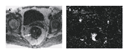
In many types of cancer, nodal disease is an independent adverse prognostic factor. However, measurement of the nodes is the only widely accepted method of assessing nodal involvement by means of imaging. The information obtained from accurate preoperative nodal staging can influence the oncologist's decision to use neoadjuvant therapy or the surgeon's decision to perform nodal dissection. Indeed, the results of preoperative nodal staging may directly determine whether radical treatment is initiated. Such an approach cannot yet be reliably implemented, since the specificity and negative predictive value of nodal staging according to size are moderate at best. Clearly, a more accurate image-based method of distinguishing malignant from nonmalignant lymph nodes is needed.
In this issue of the Journal, Harisinghani and colleagues (pages 2491�2499) report the use of lymphotropic superparamagnetic nanoparticles, a novel MRI contrast agent, in the nodal staging of prostate cancer. Seventy-one percent of malignant nodes detected with MRI with lymphotropic superparamagnetic nanoparticles were smaller than the threshold size (10 mm) used to identify nodal disease on conventional imaging. MRI with lymphotropic superparamagnetic nanoparticles had a high overall sensitivity, specificity, and accuracy on both a per-patient and a node-by-node basis. In addition, the negative predictive value of the test was impressively high. The results of the study demonstrate that imaging can be used to identify metastatic infiltration in nodes measuring 5 to 10 mm. This has important implications, because the technique may be used to select patients for extended lymphadenectomy or to delineate radiotherapy fields.
The success of a study that correlates radiological and pathological findings depends on the accuracy with which nodes visible on imaging can be matched to those in the pathological specimen. Drawing on our own studies using MRI with lymphotropic superparamagnetic nanoparticles in patients with rectal cancer, we emphasize that even with the most meticulous attention to detail, perfect matching may be impossible.
Despite the encouraging findings, the success of MRI with lymphotropic superparamagnetic nanoparticles may still be limited by the size of the involved nodes. Harisinghani et al. found that the sensitivity of this approach was substantially lower for nodes measuring less than 5 mm than for larger nodes. Furthermore, although it may be possible to use MRI with lymphotropic superparamagnetic nanoparticles to detect metastases measuring 1 to 2 mm, it is unclear whether the technique will reliably identify small metastatic foci in nodes that are partially replaced by tumor. Since an MRI examination is needed both before and after the administration of contrast medium, a cost-effectiveness analysis may be necessary to determine the appropriate clinical use of this technique. If MRI with lymphotropic superparamagnetic nanoparticles is introduced widely into clinical practice, there are also likely to be practical problems related to the performance of two MRI examinations within a 24-hour period.
There is now substantial evidence that the use of PET with fludeoxyglucose F 18 ([18F]fluoro-2-deoxy-D-glucose) can improve the accuracy of cancer staging in a cost-effective manner. The additional information provided is primarily due to the high sensitivity of the technique in detecting small-volume disease. In addition, it can be used to rule out tumors in enlarged, reactive lymph nodes. These attributes have led to improvements in both nodal and metastatic staging but overall have had little effect on tumor staging, owing to the limited spatial resolution and poor definition of adjacent landmarks, as compared with those of morphologic imaging techniques.
Combining PET with fludeoxyglucose F 18 and CT could potentially maximize the advantages of each technique while minimizing the disadvantages. Until now, the enthusiasm for combined PET�CT imaging has been based on anecdotal reports. There has been little evidence that combined PET�CT provides better results than visual correlation of PET and CT images arranged side by side or, indeed, PET alone. In this issue of the Journal, Lardinois et al. (pages 2500�2507) report that integrated PET�CT is more accurate diagnostically in the staging of non�small-cell lung cancer than PET alone, CT alone, or visual correlation of PET and CT. Tumor staging � a weak area for PET alone � and nodal staging were both significantly more accurate with integrated PET�CT, as was metastasis staging in some patients.
Integrated PET�CT could potentially reduce the number of false positive PET studies by correctly factoring in the physiological variation in uptake in a specific organ or structure. It may also be able to pinpoint areas of subcentimeter disease before biopsy or excision is performed. The role of integrated PET�CT in planning radiotherapy must also be fully explored. In this context, it could be used to delineate more accurately the areas of active tumor within an organ or, indeed, within the tumor itself. Once PET tracers that are more tumor-specific are introduced into clinical practice, integrated PET�CT will be even more important as a diagnostic tool.
Whether the proven and potential advantages of integrated PET�CT are sufficient to offset the higher capital and running costs of the combined systems has yet to be determined. Most patients who are currently referred for PET�CT have already undergone diagnostic CT. At the moment, many PET�CT units are performing simple CT studies involving low doses of ionizing radiation for the purpose of anatomical localization of PET findings alone, rather than full diagnostic CT examinations. This approach has the benefit of limiting the patient's exposure to ionizing radiation. In the future, integrated PET�CT may be routinely performed early in the diagnostic workup. The imaging approaches used by Harisinghani et al. and Lardinois et al. represent major advances in cancer imaging, which may help optimize patient care by pinpointing even the smallest tumors and providing a functional assessment of malignant disease.
Source Information
From the Academic Department of Radiology, Royal Marsden Hospital, Sutton, United Kingdom.